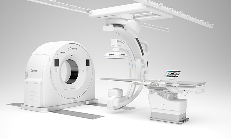
Refine and redefine intervention using our High Definition technology
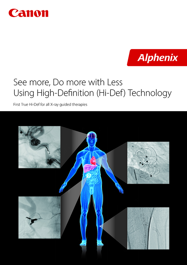
The time has come to visualize complex and intricate anatomy with a level of detail you never thought possible. Canon Medical’s Hi-Def imaging1 mode allows you to effortlessly zoom the image up to 1.5 inches at 76 micron resolution without losing image quality, helping clinicians visualize fine details, anatomical structures and deploy devices with accuracy and confidence. Discover clinical advantages now available on the world’s first 12" × 16” True Hi-Def Detector for Interventional Radiology (IR), Oncology (IO), Neurology (IN), and Cardiology (IC).
-See more, Do more with Less Using High-Definition (Hi-Def) Technology
Experience the visualization like never before with the world's first high definition mode for universal/ multi-use/interdepartmental purposes.
-High-Definition (Hi-Def) Imaging
The ability to magnify the field of view while increasing inherent spatial resolution with Hi-Def technology enhances visualization during critical aspects of endovascular interventions and has the potential to translate into additional accuracy and precision in X-ray imaging guided procedures with enhanced real time visualization during complex endovascular procedures.
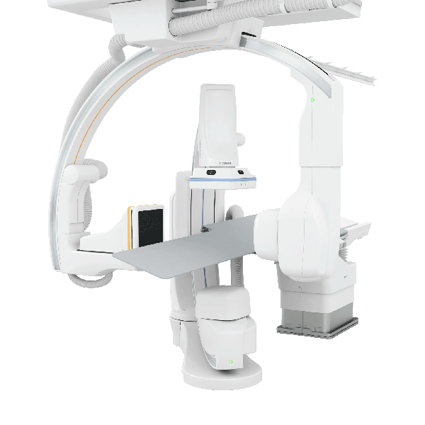
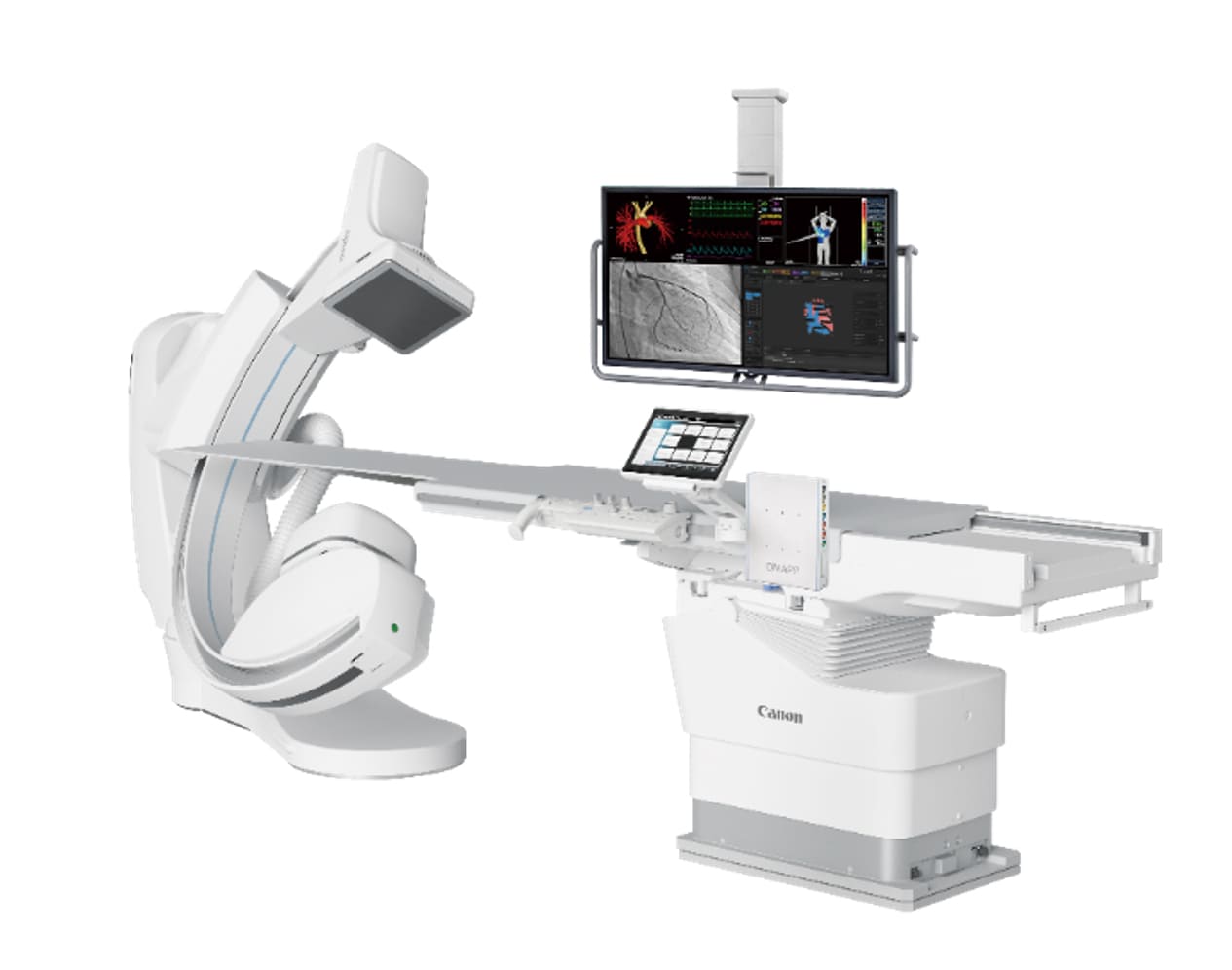
Canon Medical provides an innovative technology with the world’s first Hi-Def detector—offering more than twice the spatial resolution of conventional flat panel detectors (FPD)—for resolving fine details. This unique hybrid 12" x 12" or 12" x 16" FPD combines high definition imaging technology based on CMOS that boosts spatial resolution up to 6.6 line pairs per millimeter (lp/mm) with 76 micron pixels (Figure 1). This unique Alphenix system offers the standard magnification modes 16", 12", 10", 8", 6" or 4.3" fields of view (FOV) and three additional Hi-Def modes with 3", 2.3" and 1.5" FOV, allowing increased spatial resolution without interruption of procedure workflow. At any given point in time, both modes are available, and when needed, the selection between the two modes can be quickly changed using an FOV switch, without adding additional delay to the procedure.
-Modes Up to 2.5x Higher Spatial Resolution
Advancing minimally invasive treatments for neurovascular diseases such as stroke and aneurysms, places increasing demands for high definition real time imaging for aiding the interventionist in guiding the catheters upon identifying disease area, and to deploy treatment devices such as balloons, coils, stents, or flow diverters. Higher spatial resolution modes of the Hi-Def detector can provide sharper and visually improved images, as quantified using standard physical metrics, compared with those of traditional FPD images.1 For example, the modulation transfer functions (MTFs) of the new detector under Hi-Def mode and the FPD mode demonstrate the ability of the Hi-Def magnification modes to image spatial frequencies that would otherwise be aliased (i.e. not visualized) by the FPD magnification modes. Inevitably, Hi-Def outperforms the FPD at all spatial frequencies.
-Visualization Like Never Before
During intervention, using large FOV standard resolution FPD modes for coarse navigation, the catheter systems and devices are guided from the access site to just proximal to the lesion. Final placement and deployment of devices are performed under the magnified high-resolution Hi-Def modes with smaller FOVs (Figure 2). In a blinded-rater study that compared Hi-Def and FPD images, Hi-Def images were rated sharper and visually preferred [rated “much better” in 73% of instances] compared to the lower resolution images of the FPD.
-Case illustration2
A patient with no remarkable medical history presented to the emergency room with the worst headache of her life. Magnetic resonance angiography demonstrated a right carotid cavernous aneurysm measuring 11 × 7 mm. Lumbar puncture and head CT scan were negative for subarachnoid hemorrhage. The aneurysm size and location were confirmed with diagnostic cerebral angiography.
Under 2.3" x 2.3" FOV Hi-Def mode roadmap guidance, a 2.5Fr microcatheter over a 0.014" microwire was introduced and placed in the right distal M1 segment (Figure 3). An 2.4Fr microcatheter with a 45° angle was introduced concomitantly over the 0.014" microwire to cannulate the aneurysm neck. Under Hi-Def magnification, a single 9 × 33 mm coil was partially deployed into the aneurysm dome. Subsequently, a 4.5 × 23 mm LVIS Blue stent was introduced into the 2.5Fr microcatheter and deployment was started just distal to the aneurysm neck. Under 2.3" x 2.3" FOV Hi-Def mode, deployment showed good wall apposition throughout the curve of the cavernous carotid artery. The partially deployed coil was then fully deployed, followed by a second 5 × 20 mm coil. After the second coil deployment, DSA contrast runs were obtained and showed stasis in the aneurysm dome. The imaging mode was switched to large FOV, and a DSA contrast run was obtained to make sure the distal circulation was patent post-treatment. The patient was extubated and remained neurologically intact.
-To assess the impact of improved visualization provided by the Hi-Def mode during cerebral aneurysm treatment a Pipeline Flow Diverter, a post-procedure physician survey was conducted in a single center study. Consecutive patients over a 10-month period treated with the use of the dual resolution imaging detector were included. A summary of the responses is presented in Figure 4 from a total of 25 cases.4 In all the cases reported, 100% of the time physicians either agreed or strongly agreed that Hi-Def mode visualization during the procedure was improved compared with standard FPD.
One critical aspect of deployment of a Pipeline Flow Diverter is the visualization of the distal end of the flow diverter as it is deployed and the subsequent opening of the device. Due to lack of radial forces, the distal end of the device may not immediately open. Visibility of this detail can dictate the next steps of the deployment procedure for instance wait for the distal end to open or re-sheath and redeploy the flow diverter. The high resolution Hi-Def modes offer the interventionalist a definite advantage in accurate placement and deployment of devices.
-Redefine Lower Extremity Intervention with High Definition (Hi-Def ) Imaging
The new Alphenix family of interventional systems equipped with Hi-Def imaging helps clinicians see fine details with clarity and precision during interventional procedures