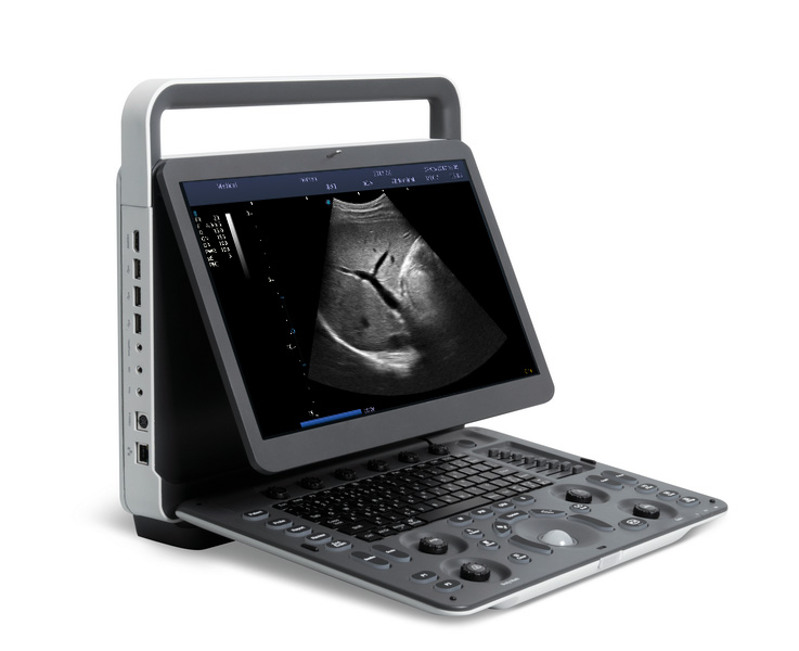
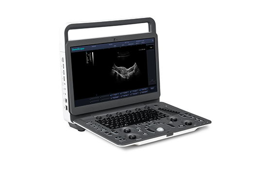
Danus 30
Ultrasound Diagnostic System
A Smart Choice for Efficient Diagnosis
Besides unique Hi platform multi beam technology and BTM transducer technology, Danus 30's compact, well-balanced design, high mobility and built-in battery make it a smart choice for doctors' daily use.
-High-definition 21.5-inch monitor
-13.3-inch touch screen
-Cable management solution
-Built-in battery for scanning
Advanced Applications
Panoramic
The extended field of view displays more image information without sacrificing image quality.
Elastography
Real time elastography is a new noninvasive and painless technique that can help determine the hardness of organs and other structures such as breast, thyroid. Elastic imaging provides users with dynamic visual information and displays the rigidity of organs, which is helpful for direct and quantitative diagnosis and treatment.
eBiopsy+
The image of the puncture needle is enhanced by the deflection of the acoustic beam, including needle enhancement, needle tip red rendering, virtual needle passage and scale line, supporting auto steering.
Tissue Doppler Imaging
Tissue Doppler Imaging (TDI) is a robust and reproducible echocardiographic tool that employs the Doppler effect to assess muscle wall characteristics throughout the cardiac cycle including velocity, displacement, deformation, and event timings. It has permitted a quantitative assessment of both global and regional function and timing of myocardial events
Curved AM
Curved Anatomical M-Mode (CAM) technology can show all the spatial and temporal relationship of myocardial segment movements during the cardiac cycle in the scanning sector, which provides a new measurement method to quantitatively analyze the abnormalities of segmental myocardial motion during systolic or diastolic period.
Smart Workflow
Auto Volume Flow
Measure the blood vessel area, the blood flow velocity could be measured by spectrum automatically, then the blood flow volume results will show.
Auto IMT
Automatic identification and measurement of intima-media thickness. Both left and right blood vessels, anterior and posterior walls can be measured.
fAssist
Providing tutorial information regarding abdomen, vascular, small parts, GYN, MSK, etc., including standard ultrasound image, anatomical diagram, scanning technique and tips.
Dual Live
Dual Live:B and B+C images are displayed in the same time for better diagnosis.
*Transducers :-
1) Convex C5-1
Applications:Abdomen,
Obstetrics, Gynecology
2) Micro-convex MC10-3
Applications:Pediatrics,
Cardiology
3) Intracavitary EC9-4
Applications:Obstetrics,
Gynecology, Urology
4) Linear L12-4
Applications:Small parts,
Vascular, MSK
5) Linear L17-5
Applications:Small parts,
Vascular, MSK
6) HD Linear L13-3
Applications:Small parts,
Vascular, MSK, Breast
7) Phased Array P5-2
Applications:Cardiology,
Abdomen, TCD
8) Phased Array P8-2
Applications:Abdomen,
Pediatric cardiology
9) Convex Volume V6-2
Applications:Abdomen,
Obstetrics, Gynecology